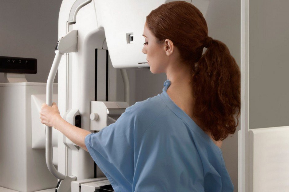
Increases Breast Cancer Mortality
Mammography is the most widely used screening modality for breast cancer and with good reason for the medical community. It gives them more patients. Breast cancer screenings result in an increase in breast cancer mortality and fail to address prevention.
Diagnosis of cancers that would otherwise never have caused symptoms or death in a woman's lifetime can expose a woman to the immediate risks of therapy (surgical deformity or toxicities from radiation therapy, hormone therapy, or chemotherapy), late sequelae (lymphedema), and late effects of therapeutic radiation (new cancers, scarring, or cardiac toxicity). Although the specific plan of oncologists is usually to recommend tailored treatments according to tumor characteristics, there is still no reliable way to distinguish which cancer would never progress in an individual patient; and consequently treatments are lumped into the "treat all just in case" just in case category.
Breast cancer screening does not play a direct role in the reductions of deaths due to breast cancer in almost every region in the world. Part of the failure correlates to more than 70 percent of mammographically detected tumors being false positives leading to unnecessary and invasive biopsies and subsequent cancer treatment such as radiation which itself causes cancer.
More Than Half Result In Overdiagnosis
Out of all breast cancers detected by screening mammograms, up to 54% are estimated to be results of overdiagnosis. The best estimations of overdiagnosis come from either long-term follow-up of RCTs of screening or the calculation of excess incidence in large screening programs.
Despite no evidence ever having supported any recommendations made for regular periodic screening and mammography at any age, malicious recommendations from the Society of Breast Imaging (SBI) and the American College of Radiology (ACR) on breast cancer screening are now promoting that breast cancer screening should begin at age 40 and earlier in high-risk patients. Published in the Journal of the American College of Radiology (JACR), the recommendations released by the SBI and ACR state that the average patient should begin annual breast cancer screening at age 40. They also target women in their 30s if they are considered "high risk" as they stated.
On average, 10% of women will be recalled from each screening examination for further testing, and only 5 of the 100 women recalled will have cancer. Approximately 50% of women screened annually for 10 years in the United States will experience a false positive, of whom 7% to 17% will have biopsies. The risk of cancer increases as much as 30% in a given 10 year period of women being exposed to yearly mammograms.
Inaccurate Even When Cancer Is Present
6% to 46% of women with invasive cancer will have negative mammograms, especially if they are young, have dense breasts, or have mucinous, lobular, or rapidly growing cancers.
Radiation-induced mutations can cause breast cancer, especially if exposure occurs before age 30 years and is at high doses, such as from mantle radiation therapy for Hodgkin disease. The breast dose associated with a typical two-view mammogram is approximately 4 mSv and extremely unlikely to cause cancer. One Sv is equivalent to 200 mammograms. Latency is at least 8 years, and the increased risk is lifelong.
The rate of advanced breast cancer for U.S. women 25 to 39 years old nearly doubled from 1976 to 2009, a difference too great to be a matter of chance.
In 1976, 1.53 out of every 100,000 American women 25 to 39 years old was diagnosed with advanced breast cancer, a study in the American Medical Association found. By 2009, the rate had almost doubled to 2.9 per 100,000 women in that age group -- a difference too large to be a chance result.
Cause Far More Harm Than Good
A disturbing study published in the New England Journal of Medicine is bringing mainstream attention to the fact that mammography has caused far more harm than good in the millions of women who have employed it over the past 30 years as their primary strategy in the fight against breast cancer
Titled "Effect of Three Decades of Screening Mammography on Breast-Cancer Incidence," researchers estimated that among women younger than 40 years of age, breast cancer was overdiagnosed, i.e. "tumors were detected on screening that would never have led to clinical symptoms," in 1.3 million U.S. women over the past 30 years. In 2008, alone, "breast cancer was overdiagnosed in more than 70,000 women; this accounted for 31% of all breast cancers diagnosed.
Most mammography-detected breast cancer presents without symptoms in the majority of women within which it is detected, and if left untreated will not progress to cause harm to women. Indeed, without x-ray diagnostic technologies, many if not most of the women diagnosed with it would never have known they had it in the first place. The journal Lancet Oncology, in fact, published a cohort study last year finding that even clinically verified "invasive" cancers appear to regress with time if left untreated:
"[We] believe many invasive breast cancers detected by repeated mammography screening do not persist to be detected by screening at the end of 6 years, suggesting that the natural course of many of the screen-detected invasive breast cancers is to spontaneously regress."
The study authors point out "The introduction of screening mammography in the United States has been associated with a doubling in the number of cases of early-stage breast cancer that are detected each year." And yet, they noted, only 6.5% of these early-stage breast cancer cases were expected to progress to advanced disease. Mammography-detected breast cancer and related 'abnormal breast findings,' in other words, may represent natural, benign variations in breast morphology. Preemptive treatment strategies, however, are still employed today as the standard of care, with mastectomy rates actually increasing since 2004.
It is also questionable whether screening mammograms can even provide genuine 'early diagnosis' as is frequently claimed. A new blood test being developed in America and Nottingham, England will pick up on proteins developed by the very earliest 'rogue' cells almost before a cancer has formed. In the press release the scientists claim that this is a good 4 years before a mammogram can show up a tumour. Apparently, a cancer makes about 40 divisions during its life, and mammograms cannot pick up a breast tumour until it is of a sufficient size, usually around 20 such divisions. So much for early diagnosis!
These concerns are part of a growing trend. Perhaps one of the most damning reports was a large scale study by Johns Hopkins published in 2008 in the prestigious Journal of the American Medical Association's Archives of Internal Medicine (Arch Intern Med. 2008;168[21:2302-2303). In the Background to the research it was pointed out that breast cancer diagnosis rates increased significantly in four Scandanavian counties after women there began receiving mammograms every two years.
The Dangers of Routine Mammography
The recent Komen controversy has the media buzzing about a reversal of policy over its decision to cut funding to PP and mammogram screening procedures. The real issue for women's health is not about funding but about the deadly effects from radiation spewing from mammogram screening devices.
Routine mammograms are far less effective at preventing breast cancer deaths and far more expected to cause unnecessary procedures, over-treatment and ultimately accelerate death more than any other screening method on women.
- A routine mammogram screening typically involves four x-rays, two per breast. This amounts to more than 150 times the amount of radiation that is used for a single chest x-ray. Bottom line: screening mammograms send a strong dose of ionizing radiation through your tissues. Any dose of ionizing radiation is capable of contributing to cancer and heart disease.
- Screening mammograms increase the risk of developing cancer in premenopausal women.
- Screening mammograms require breast tissue to be squeezed firmly between two plates. This compressive force can damage small blood vessels which can result in existing cancerous cells spreading to other areas of the body.
- Cancers that exist in pre-menopausal women with dense breast tissue and in postmenopausal women on estrogen replacement therapy are commonly undetected by screening mammograms.
- For women who have a family history of breast cancer and early onset of menstruation, the risk of being diagnosed with breast cancer with screening mammograms when no cancer actually exists can be as high as 100 percent.
Thermography (also called thermology) is a little-known technique for breast cancer detection that's been available since the 1960s. It's non-invasive and non-toxic, using an infrared camera to measure thermal emissions from the entire chest and auxiliary regions. Cancerous tissue develops a blood supply to feed a growing tumor, and the abnormal blood vessel formations generate significantly more heat than the surrounding healthy tissue. The infrared camera detects the differences in heat emitted from abnormal tissue (including malignancies, benign tumors and fibrocystic disease), as compared to normal tissue. There is no physical contact with the patient, who stands several feet away from the camera while a technician takes a series of images.
A second set of images is taken following a "cold challenge". The patient places her hands in ice cold water for one minute causing healthy tissue to constrict while the abnormal tumor tissue remains hot. The infrared scanner easily distinguishes the difference, and these images are compared with the first set for confirmation.
Thermography can detect abnormalities before the onset of a malignancy, and as early as ten years before being recognized by other procedures such as manual breast exam, mammography, ultrasound or MRI. This makes it potentially life-saving for women who are unknowingly developing abnormalities, as it can take several years for a cancerous tumor to develop and be detected by mammogram. Its accuracy is also impressive, with false negative and false positive rates at 9% for each. Thermography is also an effective way to establish a baseline for comparison with future scans; therefore, women should begin screening by the age of 25.
Comment: Iodine has been proven to both prevent and treat breast cancer.
- The Thyroid-Breast cancer connection: Iodine deficiency can lead to thyroid issues & eventually breast cancer
- Iodine treats breast cancer and more, overwhelming evidence
- Iodine deficiency linked to thyroid and breast cancer, fibrocystic breast disease, infertility, obesity, mental retardation & halide toxemia
Although widely embraced by alternative health care practitioners, thermography's obscurity in the mainstream means that too many women rely on mammograms as their only option. There are several reasons for thermography's lack of support by the conventional medical community. Early thermal scanners were not very sensitive, nor were they well-tested before being used in clinical practice. This resulted in many misdiagnosed cases and its utter dismissal by the medical community. Since then the technology has advanced dramatically and thermography now uses highly sensitive state-of-the-art infrared cameras and sophisticated computers. A wealth of clinical research attests to its high degree of sensitivity and accuracy. In 1982, the FDA approved thermography for breast cancer screening, yet most of the medical establishment is either unaware of it or still associates it with its early false start. Since most women are also uninformed of the technology there is no pressure on the medical community to support it.
Modern-day breast thermography boasts vastly improved technology and more extensive scientific clinical research.
In fact, the article references data from major peer review journals and research on more than 300,000 women who have been tested using the technology. Combined with the successes in detecting breast cancer with greater accuracy than other methods, the technology is slowly gaining ground among more progressive practitioners.
Sources:
nlm.nih.gov
cancer.gov
jamanetwork.com
ncbi.nlm.nih.gov
senkyo.co.jp
nejm.org
nap.edu
books.google


I never knew cannabis oil was indeed wonderful
and very effective in treating cancer’ if not for the
government and their so called rules in regulating
cannabis my Dad would have still been alive.
thanks to the newly policy for legalizing cannabis
in my state i would have still lost my son to
kidney cancer, i was really touched and surprised
when i watch lots of documentary on how
cannabis oil had helped lot of people whom their
family members never thought they could make it
after undergoing several ”Chemo” from the dept of
my heart i must say a word of appreciation to Mr.
Rick Simpson for the timely intervention in the life
of my son suffering from Kidney Cancer. as i am
writing this testimony on this Blog my Son is so
strong and healthy in spite he hasn’t completed
the total Dosage’ for your cannabis and medical
consultation try and get in touched with him
through his email: phoenixtears1112@gmail.com so he
can enlightened you more.