For the first time, scientists have captured detailed images of life's essence.
The dazzling pictures reveal a key step in the process of cell division, which all organisms must undergo to survive. The moment occurs deep within a cell, as two proteins work in concert to unzip a strand of DNA to create two new cells.
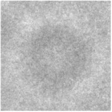
|
| ©MariaSchumacher/M.D. Anderson |
| An electron microscope image shows a complex of proteins and DNA shortly before the proteins shear the DNA into two. |
But until now, scientists seeking to directly observe this essential process only could view fuzzy images taken by an electron microscope.
A scientist at the University of Texas M.D. Anderson Cancer Center has changed that by perfecting a technique employed by biophysicist Rosalind Franklin more than half a century ago to gather the first images of DNA.
The new work, by biochemist Maria Schumacher, goes far beyond taking pretty pictures of life in motion, however.
Her efforts are part of a widespread push by biomedical scientists to understand the molecular cause of disease. By fully comprehending these root causes, they hope to devise more effective disease treatments.
"If we understand in great detail the structure of the proteins involved in normal cell growth, then we can understand when there's been a change to cause some sort of defect," said William Klein, chairman of M.D. Anderson's department of biochemistry and molecular biology.
By understanding the precise mechanism by which a cancer cell divides, for instance, it might be possible for scientists to develop a better drug to stop the process.
Schumacher is at the forefront of the growing field of structural biology, in which scientists have used sophisticated techniques to make atom-by-atom maps of proteins and, more recently, multiprotein structures.
There are hundreds to thousands of atoms in each protein, the workhorses inside human cells that fold into precise shapes as they carry out myriad tasks, including the essential chore of making copies of DNA for new cells.
A decade ago, a handful of proteins were mapped each year. Now, Schumacher said, there are about 1,000 a year.
Far trickier to map are multiprotein complexes, such as those Schumacher imaged to better understand cell division. She had to capture more than one protein at a time, freezing a moment in their delicate folding dances. It was as much art as science.
To get fine pictures of these structures, scientists must "crystallize" them, meaning they must remove all impurities and, using a variety of techniques, condense what remains behind until it becomes a solid structure. That is easier said than done. Schumacher failed 42 times before creating a suitable crystal of one particular protein. "It's frustrating sometimes," she said. "But in the end, to me, it's always worth it to see the final picture."
After crystallizing a protein or multiprotein structure, Schumacher travels to Lawrence Berkeley Laboratory in California to use its powerful synchrotron, a particle accelerator that bombards the crystals with high-intensity X-ray beams. This reveals a map of electrons a computer program can compile into the protein's structure in a process called X-ray crystallography. Schumacher's had more luck than most crystallizing proteins.
"These are really blue-ribbon structures," said Yousif Shamoo, an associate professor of Biochemistry and Cell Biology at Rice University who is familiar with Schumacher's work. "It's not like people haven't been trying to do this for years. She's just been successful at it."
Since 2001, Schumacher has had three papers published in Nature, two in Cell and one in Science, the three most exalted journals for basic science research.
In addition to having a knack for crystallizing proteins, Schumacher seems to intuitively know which protein images will reveal something about their function in addition to their structure, Klein said.
The most famous example of structure indicating function came from Franklin's crystallography images of DNA, which enabled biologists James Watson and Francis Crick to make their Nobel-winning discovery. They deduced from DNA's double-helix shape how the strands could come apart and replicate.
Schumacher's images of the heretofore-unseen machinations of cell division reveal the finer details of this earlier, landmark work. And like the first images of DNA, they reveal something about the function of proteins.
It is clear from her images that, in cell division, one protein first forms a wheel-shaped scaffold around which the DNA curls. With the DNA held into place, a second protein comes along and scissors it in two.
And life begins anew.


Reader Comments
to our Newsletter