The technique, dubbed "Brainbow" by its Harvard University inventors, is detailed in the Nov. 1 issue of the journal Nature.
|
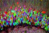
| ©Credit: Livet et al. Nature 1st Nov 2007 |
| In 'Brainbow' transgenic mice, nerve cells randomly express fluorescent proteins of different colors. Combinations of these proteins label neurons with multiple distinct hues, as seen here neurons of the hippocampus (confocal microscopy, dentate gyrus). |
Previous techniques for highlighting neurons used at most two colors. One common approach developed in 1873 by an Italian physician and still used today, called the Golgi method, stains neurons in their entirety but only affects a few brain cells at a time.
In contrast, Brainbow allows researchers to tag several hundred neurons at once with roughly 90 distinct colors. The resulting images , which resemble abstract color paintings, are both beautiful and informative. They look like they could hang in a modern art museum and are among the most detailed images of neuronal connections ever made.
As seen on TV
"We've already used Brainbow to take a first peek at the nervous system of mice, and we've observed some very interesting, and previously unrecognized, patterns of neuron arrangement," said study team member Joshua Sanes. "As far as understanding what we're seeing, we've only just scratched the surface."
To create the images , Brainbow employs a method similar to that used to generate colors on a computer or TV screen.
"In the same way that a television monitor mixes red, green and blue to depict a wide array of colors, the combination of three or more fluorescent proteins in neurons can generate many different hues," said study team member Jeff Lichtman.
But instead of red, green and blue light, Brainbow relies on cyan, red and yellow gene pigments. The red gene pigment comes from coral, while the cyan and blue pigments are modified versions of a fluorescent green pigment found in jellyfish.
Using genetic recombination techniques, the researchers bundled the pigment-expressing genes into DNA packages and inserted them into the genomes of developing mice. As the mice develop, the pigment genes get divvied up between the rodent cells. Study team member Jean Livet likens the DNA package to a "molecular slot machine."
"Each cell would play the slot machine and be attributed a different color," Livet told LiveScience.
In order for the color genes to be expressed, however, the mice cells must also contain another gene, called Cre. Derived from bacteria, Cre activates the color genes inside the cell. If the color genes are the slot machine, then Cre is "the hand pulling the lever over and over again," Livet said.
By using mice that express Cre in different parts of their bodies or at different times during development, scientists can use Brainbow on different cell types. "The system can be tuned to whatever you want," Livet said.
The entire circuit
The colors are only visible when viewed under fluorescent light, so the Brainbow-ed brains still look like normal mice brains, Livet said, "or normal transgenic mice brains, I should say."
Brainbow does have some disadvantages. For one, it relies on fluorescent microscopes, which can cost several hundred thousand dollars. "It's not like the Golgi stain, where you can just look through a normal microscope," Livet said.
Another limitation is that it only works with genetically modified, or transgenic, animals, which at the moment only include mice. With the Golgi stain, "you can do everything, including humans," Livet said.
In exchange, however, Brainbow could give neuroscientists a more complete view of the brain. "You can see how cells interact together," Livet said. "Instead of having a vision of just one cell within a circuit, you have a vision of the circuit itself."


Reader Comments
to our Newsletter