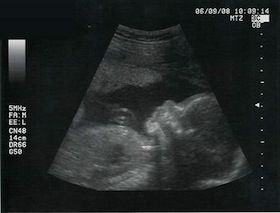
"The routine use of ultrasound in pregnancy is the biggest uncontrolled experiment in history."In the first article in this series on natural childbirth, I presented evidence that - contrary to popular belief - hospital birth is no safer than home birth.
Beverly Beech, birth activist
I'd like to begin this next article by telling you what it is not. It is not a blanket condemnation of ultrasound, nor is it a judgment of women who choose routine ultrasound during their pregnancy. It is not an argument against using ultrasound to investigate suspected problems, or to detect potential abnormalities, provided the woman is adequately informed.
The purpose of this article is to clarify the issues surrounding ultrasound's use in clinical practice, to critically examine the clinical benefit of routine prenatal ultrasound, and to raise awareness of the potential risks associated with repeated ultrasound scans.
This was going to be a very long article, so I decided to split it into two parts. In part A I will discuss the use of ultrasound in clinical practice and examine whether it improves birth outcomes. In part B, I will review studies on the safety of ultrasound as it is used today, and make recommendations for expecting mothers.
History of ultrasound and use in clinical practice
Ultrasound was originally developed in WWII to detect enemy submarines. After the war in 1955, a surgeon in Glasgow named Ian Donald began to experiment with it for medical uses. Using beefsteaks as "control" subjects, he scanned the abdominal tumors he had removed from his patients and found that different tissues gave different patterns of sound wave echo. He quickly realized the potential of ultrasound for examining a growing baby in utero.
Initially, ultrasound was used only to investigate possible problems. For example, if there was bleeding in early pregnancy, it would be used to determine whether miscarriage was inevitable. Later in pregnancy, if breech or twins were suspected, ultrasound would be used to confirm that suspicion. In these cases, ultrasound can be very useful for a woman and her caregivers.
However, over the years ultrasound has come to be used as routine scan at 18-20 weeks for all women. This is referred to as "routine prenatal ultrasound", or RPU for short. It involves scanning all pregnant women - whether a problem is suspected or not - in the hope of improving birth outcomes.
As often happens in medicine, techniques which may be of value to a small percentage of people slowly become adopted for routine use without prior study of benefits. A perfect example of this is the alarmingly common prescription of statin drugs for women, children and men without pre-existing heart disease, in spite of the fact that they've only been shown to be effective for a small segment of the population: middle-aged men with pre-existing heart disease.
The problem with this approach, of course, is that when we perform a procedure or administer a treatment to a segment of the population without properly testing it beforehand, we are essentially conducting an uncontrolled scientific experiment on that population - often without their understanding and consent. And in this case, we are performing that uncontrolled experiment on two of the most vulnerable populations: pregnant women and babies in the womb.
Some physicians and researchers have been questioning the wisdom of performing such an experiment for decades. In 1987, UK radiologist H.B. Meire remarked:
The casual observer might be forgiven for wondering why the medical profession is now involved in the wholesale examination of pregnant patients with machines emanating vastly different powers of energy which is not proven to be harmless to obtain information which is not proven to be of any clinical value by operators who are not certified as competent to perform the operations.More recently, in 2010, the prestigious Cochrane Collaboration reviewed the available evidence on routine prenatal ultrasound (RPU) and concluded:
Existing evidence does not provide conclusive evidence that the use of routine umbilical artery Doppler ultrasound, or combination of umbilical and uterine artery Doppler ultrasound in low-risk or unselected populations benefits either mother or baby.Despite the lack of evidence supporting RPU's use in clinical practice, ultrasound is almost universally seen as a safe and effective procedure, and scans have become a "rite of passage" (in the words of Sarah Buckley) for pregnant women in most developed countries.
In the U.S., an estimated 65 to 70 percent of pregnant women have a formal scan in a diagnostic clinic, and many more women are scanned by their OB/GYN as part of their pregnancy visit.
Is ultrasound as effective and safe as we've been led to believe?
In order to answer that question, we have to distinguish between different uses of ultrasound. As I said earlier, ultrasound scanning can be a useful diagnostic tool when abnormalities are suspected. I have no argument with using it in this manner. The question I'd like to investigate here is whether routine prenatal ultrasound - when no abnormalities are suspected - is necessary and effective.
RPU is used today for several reasons:
- To predict the birth due date
- To determine the sex of the baby
- To detect potential abnormalities
- To identify placenta previa (low lying placenta)
- To assess specific markers, such as the length of woman's cervix and the amount of amniotic fluid at the end of pregnancy
"Before the development of prenatal testing for fetal abnormality the fetus was assumed to be healthy, unless there was evidence to the contrary. The presence of prenatal testing and monitoring shifts the balance towards having to prove the health or normality of a fetus."The important question is: is RPU necessary and effective for these uses? Does it improve specific birth outcomes like perinatal mortality or morbidity?
Routine prenatal ultrasound is not recommended by researchers and major organizations
In general, RPU is accurate for predicting birth date when scans are performed in the early stages of pregnancy. The estimated due date (EDD) calculated by a scan at 7-8 weeks will be accurate to plus or minus 3-4 days.
However, calculations of EDD based on a woman's menstrual cycle can be just as accurate.
What about detecting abnormalities? Studies show that RPU detects between 35-80% of the 1 in 50 babies that have significant abnormalities at birth. The larger centers with better trained sonographers have rates toward the higher end of the scale, but even major centers miss 40% of abnormalities.
That's because many abnormalities are difficult or impossible to detect with RPU. Heart and kidney problems are unlikely to be picked up, as are some markers for Down syndrome. Cerebral palsy, autism, and other markers of intellectual disability are impossible to detect.
Then there's the small but significant chance that an abnormal finding may be a false positive. A UK survey showed that for 1 in 200 babies aborted for supposed major abnormalities, the diagnosis on post-mortem was less severe than predicted by ultrasound, and the termination was probably unjustified. In the same survey, 2.4 percent of babies diagnosed with major malformations - but not aborted - had conditions that were significantly over- or underdiagnosed.
Two other studies have shown false positive results in roughly 10% of babies diagnosed with structural abnormalities. And in some cases, the abnormalities spontaneously resolve without intervention.
In addition to false positives, there are also cases that are difficult to interpret, and the outcome for the baby is unknown. This uncertainty can cause considerable stress and anxiety for the mother, which in turn adversely affects the developing baby. In one study involving women at higher risk, a full 10 percent of scans were uncertain. And in that same study, mothers with uncertain diagnoses were still anxious three months after the birth of their baby.
Ultrasound scanning for placenta previa is mostly accurate, but almost all women who test positive for it on a scan will be unnecessarily worried. Studies show that the placenta will move up and not cause problems during birth for 80 to 100 percent of women, and that detection of placenta previa by RPU is not safer than detection during labor.
All of this might explain why organizations like the American College of Obstetricians and Gynecologists recommend scans only for specific reasons, including uncertain due dates and fetal assessment, and advises that routine prenatal scans are cost-effective only when done by ultrasound technicians working in high-level centers.
In Canada, practice guidelines recommend only a single midpregnancy scan and stress that information on risks and benefits must be provided and informed consent obtained.
Routine prenatal ultrasound does not improve birth outcomes
Studies on RPU over the years have consistently shown that it does not improve birth outcomes as measured by clinical endpoints such as perinatal mortality and morbidity.
A 1993 meta-analysis of all randomized trials prior to that date covering 16,000 births showed no improvement in the condition of babies measured by APGAR score when ultrasound was used compared to those who did not have it. There was a slight reduction in perinatal mortality in this study. However, this happened because these babies were aborted during pregnancy - not because their lives were saved. There was no increase in the number of live, healthy births from RPU.
The authors of this study concluded:
Routine ultrasound scanning does not improve the outcome of pregnancy in terms of an increased number of live births or of reduced perinatal morbidity. Routine ultrasound scanning may be effective and useful as a screening for malformation. Its use for this purpose, however, should be made explicit and take into account the risk of false positive diagnosis in addition to ethical issues.In another 1993 review covering 15,530 births the authors found "no significant differences in maternal outcomes". The rates of induced abortion, amniocentesis, tests of fetal well-being, external version, induction, and cesarean section and the distribution of total hospital days were similar in the two groups. They concluded:
Screening ultrasonography resulted in no clinically significant benefit.In the same year (1993), the World Health Organization (WHO) issued a letter reviewing the studies performed on routine ultrasound to date and concluded 2:
It is fair to say that at the moment the best research shows no benefit from routine ultrasound scanning and the real possibility of serious risk. ...we urge you to reconsider all present policy with regard to routine ultrasound scanning during pregnancy, based on these important scientific papers.1. Beech, BL. Ultrasound unsound? Association for Improvements in the Maternity Services. 1996
2. Beech, BL. Ultrasound unsound? Association for Improvements in the Maternity Services. 1996.


Reader Comments
to our Newsletter